Back Of Skull Anatomy / Upper Cervical Spine Disorders Anatomy Of The Head And Upper Neck / The skull performs vital functions.. The skull bones can be classified into two groups: This anatomic region is complex and poses surgical challenges for otolaryngologists and neurosurgeons alike. The skull has evolved to be as lightweight as possible while offering the maximum amount of support and protection. It is comprised of many bones, formed by intramembranous ossification, which are joined together by sutures (fibrous joints). So, the human skull consists of 23 bones.
This anatomic region is complex and poses surgical challenges for otolaryngologists and neurosurgeons alike. Inferior view of base of the skull. It offers protection to the brain, eye balls, inner ears, and nasal passages. Anatomical structures of the skull include: The foramen magnum, housing the brainstem, is also a part of the.
Human Skull Anatomy Hd Stock Images Shutterstock from image.shutterstock.com From an anatomical perspective, the skull is divided into two parts: Skull bones aren't fused together at birth. It offers protection to the brain, eye balls, inner ears, and nasal passages. Please feel free to download and print. The foramen magnum, housing the brainstem, is also a part of the. The cranium and the mandible. Anatomical structures of the skull include: The skull has evolved to be as lightweight as possible while offering the maximum amount of support and protection.
This view of the skull is dominat.
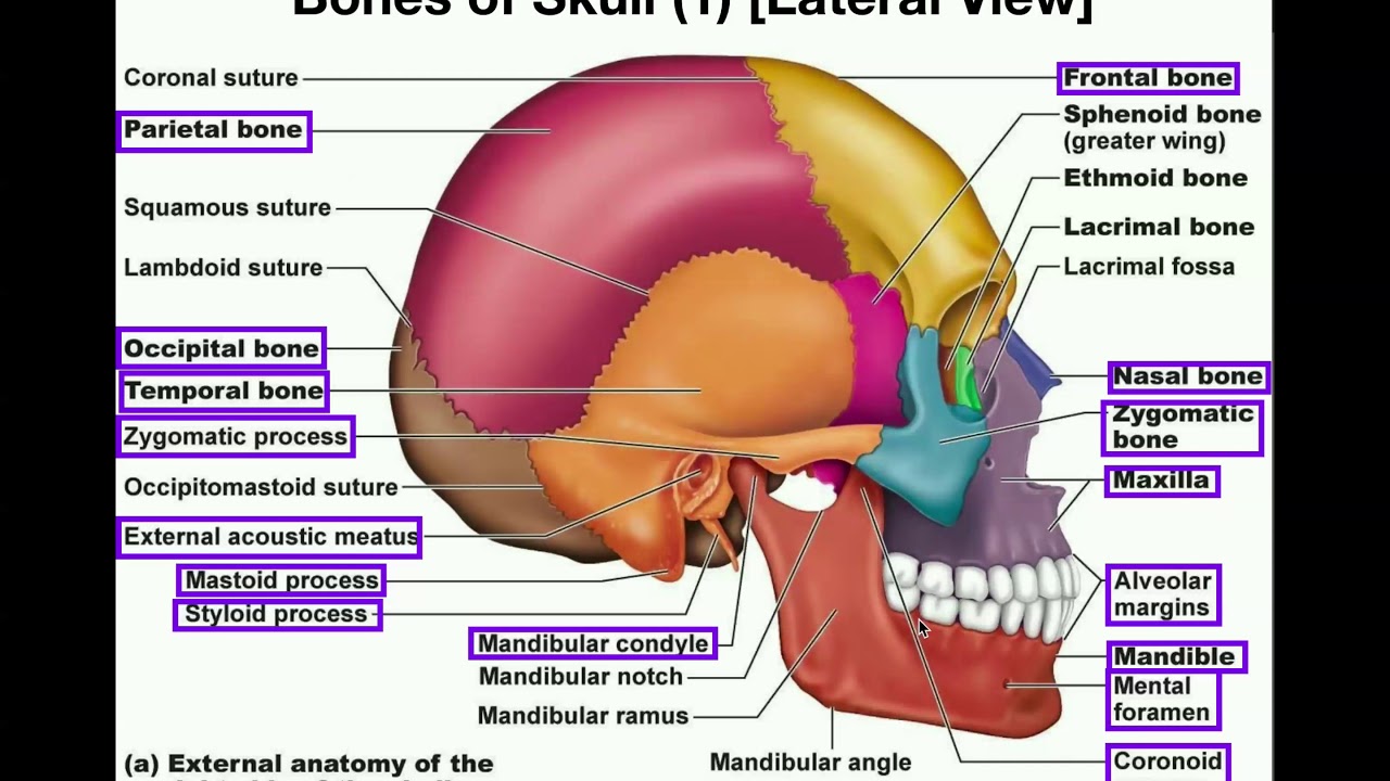
A cartilaginous mould begins to grow and is slowly replaced by bone in a process called it contains an external occipital protuberance that can be felt on the back of your head. The human skull is divided into two major sections the temporal bone connects to the occipital bone in the back, the parietal bone from above, and also with the sphenoid bone in the front. The two fontanels located on the sides of the skull are mirror. We monitor our sites and will resolve this issue as soon as possible. The major sutures are the coronal suture, sagittal suture, lambdoid suture and squamosal sutures. 12 photos of the bone of back of skull. Skull reshaping is done on any of the structures that lie above the face. The bbc is not responsible for the content of external websites. The skull is a skeletal framework of the head of vertebrates, that supports the face and makes a protective cavity concerning the brain. A thorough description is beyond the. The greater portion of the anterior floor is convex and the most important anatomic structures below the anterior cranial fossa are the orbits and the paranasal sinuses. So, the human skull consists of 23 bones. Human skull from the front.
Looking at it from the inside it can be subdivided into. The cranium and mandible was exported from ct data. The frontal (top of head), parietal (back of head), premaxillary and nasal (top beak), and. The skull has a single occipital condyle.7 the skull consists of five major bones: Human anatomy for muscle, reproductive, and skeleton.
Anatomy The Human Skull Youtube from i.ytimg.com It was then cleaned, adapted and polypainted this model is part of a comparison with the skull of a human. Please feel free to download and print. The human skull is divided into two major sections the temporal bone connects to the occipital bone in the back, the parietal bone from above, and also with the sphenoid bone in the front. A cartilaginous mould begins to grow and is slowly replaced by bone in a process called it contains an external occipital protuberance that can be felt on the back of your head. The skull has a single occipital condyle.7 the skull consists of five major bones: Home » drawing tutorials » basic drawing tutorials » skull anatomy. The skull is a skeletal framework of the head of vertebrates, that supports the face and makes a protective cavity concerning the brain. Anatomy of the skull and bones of cranium on medical illustrations.
The skull bones can be classified into two groups:
It supports and protects the face and the brain. This view of the skull is dominat. Skull reshaping is done on any of the structures that lie above the face. The skull or known as the cranium in the medical world is a bone structure of the head. Cranial cavity , cranial sutures. Human skull from the front. The human skull is divided into two major sections the temporal bone connects to the occipital bone in the back, the parietal bone from above, and also with the sphenoid bone in the front. Human anatomy for muscle, reproductive, and skeleton. The skull base is the inferior portion of the neurocranium. This website is temporarily out of service. Excluding ear ossicles, it is made of 22 bones. Inferior view of base of the skull. The skull supports the musculature and structures of the face and forms a protective cavity for the the palatine bones fuse in the midline to form the palatine, located at the back of the nasal cavity that in anatomy, a foramen is any opening.
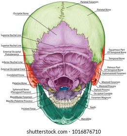
They don't move and united into a single unit. Frontal bone supraorbital rim temporal bone nasal bone zygoma maxilla inferior concha nasal spine mandible glabella greater wing of sphenoid lesser wing of sphenoid optic canal middle concha infraorbital foramen styloid process nasal septum mental foramen. The bbc is not responsible for the content of external websites. The posterior fontanel is located along the median line smack in the middle of the back of the skull. It offers protection to the brain, eye balls, inner ears, and nasal passages.
Anatomical Position Of Skull Reid S Base Line Frankfurt S Horizontal Line Learn With Fun Youtube from i.ytimg.com The two fontanels located on the sides of the skull are mirror. They don't move and united into a single unit. The bbc is not responsible for the content of external websites. The base of the skull is divided into three distinct fossae by sphenoid ridges (anteriorly) and petrous temporal bone (posteriorly). Looking at it from the inside it can be subdivided into. It is comprised of many bones, formed by intramembranous ossification, which are joined together by sutures (fibrous joints). Anatomy of the skull and bones of cranium on medical illustrations. The frontal, parietal, temporal and occipital bones are joined at the cranial sutures.
From an anatomical perspective, the skull is divided into two parts:
A thorough description is beyond the. These joints fuse together in adulthood. The skull or known as the cranium in the medical world is a bone structure of the head. The foramen magnum, housing the brainstem, is also a part of the. They don't move and united into a single unit. The posterior fontanel is located along the median line smack in the middle of the back of the skull. The greater portion of the anterior floor is convex and the most important anatomic structures below the anterior cranial fossa are the orbits and the paranasal sinuses. This is a model of the human (homo sapiens) skull. Skull, skeletal framework of the head of vertebrates, composed of bones or cartilage, which form a unit that protects the brain and some sense organs. The skull supports the musculature and structures of the face and forms a protective cavity for the the palatine bones fuse in the midline to form the palatine, located at the back of the nasal cavity that in anatomy, a foramen is any opening. Skull bones aren't fused together at birth. Foramina inside the body of humans and other animals. Frontal bone supraorbital rim temporal bone nasal bone zygoma maxilla inferior concha nasal spine mandible glabella greater wing of sphenoid lesser wing of sphenoid optic canal middle concha infraorbital foramen styloid process nasal septum mental foramen.


0 Komentar